How doctors diagnose yeast infection?
The clinical diagnosis of yeast infections is largely based on the knowledge of the types of yeast present in human beings, prone groups, the sites these yeasts target, infections caused and signs and symptoms with which patients present to the doctors. Before we can move on to the diagnosis of yeast infections, it would be better if we know some basics here.
Yeast Infection is the most common infection of fungi caused by Candida Albicans. This fungus grows on moist places thus infecting moist surfaces of body. It is Dimorphic (having two physical forms) in nature. These forms are:
- Budding Yeast form: This form is present normally in human respiratory and female genital tracts. It is non infectious and does not cause disease. It appears as round to oval yeast cells with single buds (daughter cells).
- Psuedohyphae form: It is the infectious form of yeast. It forms Pseudohyphae. Psuedohyphae are thread like structures produced by dividing yeast cells joined to each other end to end and forming branches at budding (cell dividing) points.
Who is more prone to yeast infections?
Yeast is an opportunistic pathogen i.e. it causes disease only when conditions are favorable for its growth, which grows and causes infection in places that are moist and have high pH value (alkaline). Groups most prone to yeast infections include:
- Children and young people
- Old age people
- Immuno-compromised (reduced immunity) patients e.g. patients suffering from AIDS.
- Patients undergoing some immunosuppressant therapies like chemotherapy.
- Diabetic patients.
- Females taking excessive antibiotics are more prone to develop these infections because use of antibiotics disturb normal vaginal flora, promoting the growth of opportunistic yeast.
- Serious Blood yeast infections are common in people that make use of needles and piercing instruments e.g. catheters and unsterilized syringes.
Which sites are commonly affected?
Yeast causes infections of:
- Female genitals.
- Oral cavity (thrush)
- Skin
- Male genitals
- Blood (systemic Infection)
- Nails
How doctors diagnose yeast infections?
Now that we know which yeast cause infections, the prone groups and commonly affected body sites, we move on to our main topic. Following are the main methods used by doctors for the diagnosis of yeast infection:
Evaluation of clinical symptoms
This method makes use of the identification of characteristic signs and symptoms of yeast infections. These signs and symptoms vary according to the site of infection.
In case of oral infections the following symptoms are seen upon physical examination of the patients:
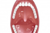
- Psuedomembrane in oral cavity is one of the most important diagnostic features. Psuedomembrane is yellowish or grayish thick adherent exudate on mucosal surfaces like tongue, throat and tonisllar pillars.
- Sore throat is present.
- There's difficulty in swallowing.
- Tongue and lips maybe swollen.
- There's formation of red and white spots around lips and margins especially near they meet (angle of the mouth).
In case of vaginal infection following symptoms are seen upon physical examination:
- There is usually a secretion of thick, foul smelling and cloudy fluid from vagina.
- There is constant itching with redness at the site of infection.
- Intercourse is painful in vagina yeast infection.
In case of skin yeast infections following symptoms are present:
- The skin becomes red.
- The skin starts to peel off.
- There is intense itching and repeated itching may lead to the ulceration of the skin.
- The affected area becomes swollen.
In male genital infections the following symptoms might be seen:
- The penis is red and swollen.
- Penis is inflamed and itchy near tip.
- Patient might also complain of pain.
In case of the infection of nails, the nails become thick, disfigured, might undergo change of color (might become white or greenish white) and may wear off in severe, prolonged infections.
In children when diapers are not changed on time yeast infection results in rash characterized by sore, swollen and red skin.
Blood infections by yeast are serious and result in inflammation and disease of heart resulting in its dysfunction. This can be diagnosed by the murmurs of heart.
Laboratory Diagnosis
If the clinical picture is not complete and further confirmation is needed, then doctors go for laboratory diagnosis. It is done by taking patient samples and diagnosing them inside laboratory by different techniques. These techniques include:
- Microscopic examination: In this methods sample is taken from infected part of the body on a sterile (germ free) cotton swab by rubbing it gently over the infected part. These samples might include skin and nail in case of skin infections and blood in case of systemic infections. Then a slide is made by putting some of this material on glass slide. 10% Potassium Hydroxide (a solution to dissolve everything except yeast cells) is put on it. Then slide is observed under microscope. Characteristic Pseudohyphae are seen. Pseudohyphae are oval Yeast cells arranged in form of long threads and branching at points of budding (division of cells).
- Fluorescent Microscopy: In this technique special kind of microscopes are used. Calcofluor-White stain is used to color Yeast cells shiny white. Fluorescent microscopes make use of special glass lens that blocks rest of the light other than stain (Polaroid) making background black. The yeast cells can then be easily seen as shiny, white colored threads branching and having oval buds (Psuedohyphae).
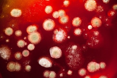
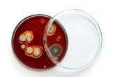
- Culturing: Yeast cells can be cultured (grown in laboratory) on special media for their diagnosis as specific organism grow in specific medium in specific form. These media have special nutrients that promote the growth of one organism while inhibiting growth of the rest. Sample is collected in form of a sterile cotton swab from infected area or nail scraping or blood. It is then introduced on special medium (for yeast it is Sabourad's dextrose agar) and left at 37 degrees for few days. White colored round colonies are formed which is diagnostic for yeast infections.
- Serological Testing: It is a method in which presence of response of body against yeast infection is measured. Body for its protection normally forms chemicals (antibodies) in response to yeast infections. These chemicals (antibodies) remain in blood. A sample is taken from blood and assessed by special laboratory machines and techniques known as ELISA (enzyme linked immunosorbent assay). A positive result may indicate previous infection thus giving false positive results.
This testing is of little use as such. However, this method is used to see if person's immunity is working or not. Small amount of non infectious material from yeast is introduced into the skin. If person has normal immunity, body will form normal amount of antibodies against yeast material which is then detected as a positive result. If result is negative immunity is compromised.
| Written by: | Michal Vilímovský (EN) |
|---|---|
| Education: | Physician |
| Published: | January 23, 2014 at 7:02 AM |
| Next scheduled update: | January 23, 2016 at 7:02 AM |
Related articles
Get more articles like this in your inbox
Sign up for our daily mail and get the best evidence based health, nutrition and beauty articles on the web.


Ache in left arm that you should not ignore
Alkaline water dangers: why you should not drink it
How to Avoid Sleepiness While Studying?
23 Foods That Increase Leptin Sensitivity
Low dopamine (e.g. dopamine deficiency): causes, symptoms, diagnosis and treatment options
Swollen taste buds: the ultimate guide to causes, symptoms and treatment
Thin endometrial lining: causes, symptoms, diagnosis and treatment
Pimples inside nose: the complete guide
Holes in tonsils: definition, symptoms, treatment and prevention
How to deal with an ingrown hair cyst
Allegra vs. Zyrtec vs. Claritin
How to get rid of phlegm (excessive mucus) in throat? Detailed guide to medical and home remedies, symptoms and causes
What causes stomach ache after meals?
Allergy to penicillin and alternative antibiotics
Liver blood test results explained